A Comprehensive Guide Through The Root Canal X-ray Procedure
Practical Tips on Preparing for a Root Canal X-Ray
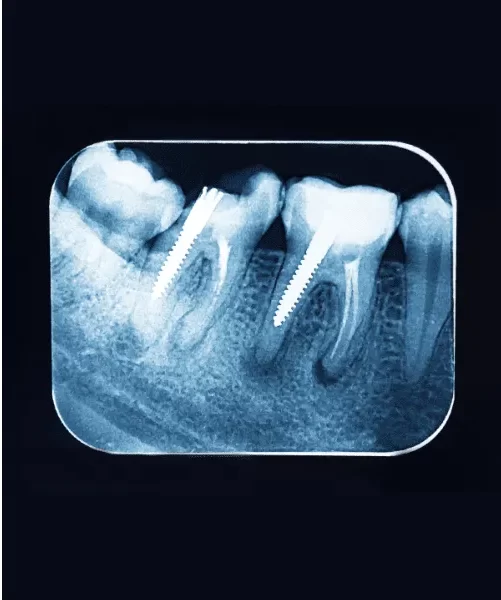
Root canal X-rays procedure are a crucial part of the diagnosis and treatment process for various dental issues. Preparing for the X-ray can help ensure a smooth experience and provide the most accurate results. Here’s what you can do to get ready, what to expect during the procedure, and any follow-up care you might need.
1. Before the X-Ray: How to Prepare for root canal x-ray procedure
- Communicate with Your Dentist:
- Let your dentist know about any medical conditions, allergies, or if you are pregnant. Pregnant patients should inform their dentist, as certain precautions, such as the use of lead aprons, will be necessary to protect the fetus.
- Avoid Eating Right Before the Appointment:
- Some patients, especially those with a strong gag reflex, may find it helpful to avoid eating for a few hours before the X-ray. This can reduce the likelihood of discomfort during the procedure.
- Practice Good Oral Hygiene:
- Brush and floss your teeth thoroughly before the appointment. A clean mouth ensures clearer X-ray images and a more comfortable experience during the procedure.
- Wear Comfortable Clothing:
- Dress in comfortable clothing, especially around the neck and shoulders, as you may need to wear a lead apron during the X-ray. Avoid wearing jewelry, especially necklaces or earrings, that could interfere with the X-ray images.
- Consider Your Comfort Needs:
- If you have a small mouth, gag reflex, or find it difficult to hold still, discuss these concerns with your dentist ahead of time. They may be able to use smaller sensors or recommend relaxation techniques to make the process more comfortable.
2. During the X-Ray: What to Expect
- Positioning:
- You will be seated in a dental chair, and your dentist or dental assistant will position you for the X-ray. They may ask you to bite down on a small sensor or film holder, which will be placed inside your mouth. The sensor may feel slightly uncomfortable, but it should only be in place for a few moments.
- Lead Apron and Thyroid Collar:
- To protect your body from unnecessary radiation, you will likely be draped with a lead apron and, in some cases, a thyroid collar. These shields are designed to block radiation from reaching other parts of your body.
- Brief and Painless Process:
- The actual X-ray exposure takes just a few seconds. You’ll need to remain still while the X-ray is being taken to ensure a clear image. The entire process is painless, though the sensor or film holder may cause mild discomfort.
- X-ray Machine Setup:
- Intraoral X-rays: For periapical views, an intraoral X-ray machine is used. The X-ray device is positioned inside the mouth to capture images of individual teeth.
- Bitewing X-rays: These views, focusing on the upper and lower back teeth, involve the patient biting down on a special X-ray film holder.
- Panoramic or CBCT X-rays: These provide a broader view and are captured with the patient’s head positioned in a specific alignment within the X-ray machine.
- X-ray Capture:
- Intraoral X-rays: The X-ray machine is activated for a brief moment, and the image receptor or film captures the X-rays that pass through the teeth.
- Bitewing X-rays: The patient holds the film holder in place, and X-rays are directed through the teeth onto the film.
- Panoramic or CBCT X-rays: The machine rotates around the head, capturing images from various angles to create a comprehensive view.
- Image Processing:
- Traditional X-rays: The captured X-ray images are processed and developed using traditional film-based methods.
- Digital X-rays: In modern practices, digital sensors or phosphor plates are used, and the images are processed digitally for immediate viewing.
- Multiple Images:
- Depending on the complexity of your case, your dentist may need to take multiple X-rays from different angles. This ensures a comprehensive view of the tooth and surrounding structures.
- Review and Analysis:
- Dentist’s Evaluation: The dentist reviews the X-ray images to assess tooth structures, root canals, surrounding bone, and any potential issues.
- Discussion with Patient: The dentist discusses the findings with the patient, explaining any identified concerns and the recommended treatment plan.
3. After the X-Ray: Follow-up Care
- No Special Care Needed:
- Typically, no special care is needed after a root canal X-ray. You can return to your normal activities immediately after the procedure.
- Discuss Results with Your Dentist:
- Your dentist will review the X-ray images with you, explaining any findings and discussing the next steps in your treatment plan. This might include a root canal procedure, further diagnostic tests, or other dental treatments.
- Address Any Discomfort:
- If the X-ray process caused any mild discomfort or soreness in your mouth, it should subside quickly. If you experience any lingering discomfort, discuss it with your dentist.
- Regular Dental Check-Ups:
- Regular dental visits and routine X-rays (as recommended by your dentist) help maintain oral health and detect any issues early. This is particularly important if you have had root canal therapy, as follow-up X-rays may be needed to monitor the healing process.
Preparing for a root canal X-ray is straightforward, and understanding what to expect can help make the experience stress-free. By following these practical tips, you can ensure a smooth process, receive accurate results, and continue with any necessary dental care promptly. Your dentist is there to guide you through each step, so don’t hesitate to ask questions or express any concerns you might have.